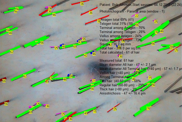
Presently the method of Phototrichogram is widely recognized and distributed in Trichology clinical practice for being highly accurate and affordable. At early stages of the disease this method helps to reveal the subclinical forms of Alopecia Androgenetica (AGA), to conduct the differential diagnosis between AGA and diffuse Chronic Telogen Effluvium (CTE), to evaluate the efficiency of the dynamic treatment regime of alopecia and much more.
Phototrichogram method was first introduced in 1970. For higher study accuracy Van Neste proposed the use of contrast enhancement of the site. R. Hoffman perfected the technique of the study conduction by introducing computer data processing. In order to start conducting a Phototrichogram study, the site for subsequent measurements shall be selected. In case if the differential diagnosis between AGA and CTE shall be established, the Phototrichogram shall be performed in two scalp areas - the frontal-parietal and occipital. In case of evaluation of the efficiency of the dynamic treatment regime of alopecia it is advisable to study the area of pronounced hair thinning or on the boundary line between affected and healthy areas. Also the standard assessment points may be used - the first point is at a distance of 2 cm from the front and 2 cm from the median line of the head, the second - at a distance of 2 cm lateral to the occiput. The hairs at selected site shall be trimmed down to minimal length by means of special portable trimmer. The size of trimmed section depends on the applied diagnostic optical equipment, it normally measures 0.8-1.5 cm in diameter. As finished it is necessary to evaluate the correctness of performed shaving - the " length uniformity" of all trimmed hairs. All hairs should be shaved down to the identical lengths! Next you have to disinfect the shaved area with some antiseptic solution and by means of a syringe with insulin needle 30-32 g or a tattoo apparatus and a tattoo or permanent make-up paint apply a dot tattoo, basically invisible by bare eye, in the center of the trimmed site, as it allows to come back in time to the same site for evaluation even in years, if needed. After 48-72 hours assess the quality of tattoo, if necessary, make a correction. Dilute a special paint for eyebrows and eyelashes to a creamy consistency and apply it to the trimmed site for all hairs to become highly visible in order to not miss any during assessment. Leave it for 10 minutes and rinse off with alcohol solution. Apply to prepared site a drop of viscous clear gel or mineral oil to be used as a contact medium. Cover the site with a coverslip glass, silica is preferable, in order to protect from any medium getting into Trichoscope lens, as well as to flatten all hairs to the scalp during diagnostic session at the same time. Next put the Trichoscope lens on it, make an image and proceed with TrichoSciencePro © computer program evaluation. The program will calculate the total number of hairs per square centimeter, as well as the number of Terminal and Vellus-like, Anagen and Telogen hairs, it will allow to determine important diagnostic features of the predominance of Vellus-like Telogen hair, average rates of hair growth and shedding, etc.
For more Phototrichogram module features of the TrichoSciencePro © computer program refer to Chapter 13. "Phototrichogram module" of the User Manual.
In case of any recent updates, for some additional information please also refer to the publication for the New Version.
For detailed Phototrichogram technique conduction in accordance to the description above refer to step by step video instructions below: